How AI in Telehealth Diagnosis Enhances Remote Healthcare

How AI in Telehealth Diagnosis Enhances Remote Healthcare

AI Health Tech Med Tech

With 76% of U.S. hospitals using telehealth services, AI plays a big role in improving diagnostic accuracy and patient care. In fact, the U.S. telehealth market is expected to reach a value of $590.6 billion by 2032. AI in telehealth diagnosis is a major factor in this surge.

Source: Tateeda

Let’s explore how AI is enhancing medical diagnosis in telehealth, and its applications.

Contents

Applications of AI in Telehealth Diagnosis

AI in healthcare

AI refers to algorithms (computer systems) that can perform tasks that typically require human intelligence. In healthcare, AI encompasses a wide range of technologies designed to assist medical professionals in various aspects of patient care (Davenport & Kalakota, 2019). These applications include:

AI’s ability to process vast amounts of data quickly and identify patterns makes it an invaluable tool in the medical field, where precision and speed can make a significant difference in patient outcomes.

How AI integrates with telehealth platforms

Telehealth platforms are increasingly incorporating AI technologies to enhance their capabilities. This integration allows for more sophisticated remote healthcare services. Here’s how AI typically works within a telehealth system:

  1. Data collection: AI systems gather patient information from various sources, including electronic health records (EHR), wearable devices, and patient-reported symptoms.
  1. Analysis: Advanced algorithms process this data to identify potential health issues or risks.
  1. Decision support: AI provides healthcare providers with insights and recommendations to aid in diagnosis and treatment planning.
  1. Patient interaction: Some AI systems can directly interact with patients through chatbots or virtual assistants, offering health advice and virtual triage services.

Key benefits of AI-powered diagnosis in telehealth

Incorporating AI into telehealth diagnosis offers several advantages:

  • Faster diagnoses: By automating certain aspects of the diagnostic process, AI can help healthcare providers reach conclusions more rapidly.
  • Cost-effectiveness: Telehealth can be cost-effective for both healthcare providers and patients. It reduces overhead costs for healthcare facilities, and lowers patient expenses related to transportation and time off work.

  • Increased accessibility: AI-powered telehealth services can extend quality healthcare to underserved areas where specialist expertise may be limited.
  • Consistency: AI systems can provide consistent analysis and recommendations, promoting similar diagnoses from different healthcare providers.

Hah & Goldin (2022) looked at how doctors use different types of patient information, especially in telehealth settings, to see where AI could help doctors manage complex patient information. As telehealth grows, doctors need to be able to make diagnoses using digital information. However, the increasing amount of patient data from mobile devices can be overwhelming for doctors.

They recommend that AI developers understand how doctors process information to create better AI tools. They also suggest that doctors should receive training in managing multimedia information as part of their education.

The Patient Experience with AI-Driven Telehealth

Now that we understand AI’s role in telehealth, it’s important to consider how these advances affect patients directly.

Hand holding phone with AI health chatbot conversation

Appointment and medication reminders

AI–powered chatbots and virtual assistants can help patients schedule and remember their doctor appointments. They can also remind patients when to take their medicines or other intermittent care they otherwise may forget.

User-friendly interfaces for remote consultations

AI is helping to create more intuitive and user-friendly interfaces for telehealth platforms. These interfaces often include:

  • Chatbots for initial patient intake and triage

  • Voice-activated assistants for hands-free interaction

  • Simplified data input methods for patients to report symptoms

Research has shown that well-designed AI interfaces can improve patient engagement and satisfaction with telehealth services.

Personalized care recommendations

AI systems can analyze individual patient data to provide personalized care recommendations. This may include:

  • Tailored treatment plans based on a patient’s medical history and genetic profile

  • Personalized medication dosage recommendations

  • Lifestyle and diet suggestions based on a patient’s specific health conditions

AI health coaching can significantly improve outcomes for patients with chronic conditions.

24/7 availability of AI-powered diagnostic tools

One of the key advantages of AI in telehealth is its ability to provide round-the-clock access to diagnostic tools. This includes:

  • Symptom checkers that patients can use at any time

  • Automated triage systems to direct patients to appropriate care levels

  • Continuous monitoring of patient data from wearable devices

Research proves that AI health services available 24/7 help treat problems earlier, particularly for patients chronic conditions that require timely treatment.

Current AI Technologies in Telehealth Diagnosis

Now that we understand how AI in telehealth improves patient engagement, let’s look at the specific technologies making this possible.

Machine learning algorithms for symptom analysis

Machine learning (ML), a subset of AI, is playing a crucial role in telehealth diagnosis through symptom analysis. These algorithms can:

  • Process patient-reported symptoms and medical histories

  • Compare symptoms against vast databases of medical knowledge

  • Suggest potential diagnoses or areas for further investigation

For example, a study published in Nature Medicine showed that an ML model can accurately diagnose common childhood diseases based on symptoms and patient history (Liang et al., 2019).

As of Fall 2023, the Food and Drug Administration (FDA) approved 692 AI or ML medical devices (531 in radiology, 71 in cardiology and 20 in neurology).

Computer vision in dermatological assessments

Tele-dermatology is another application where AI can help with remote diagnosis. Computer vision (CV) technology is making significant strides in dermatological diagnoses through telehealth. Here’s how it works:

  1. Patients upload images of skin conditions through a telehealth platform.

  2. AI-powered computer vision analyzes the images, considering factors like color, texture, and shape.

  3. The system compares the images against a database of known skin conditions.

  4. Healthcare providers receive an analysis to aid in their diagnosis.

Some AI systems can match or even exceed dermatologists in accurately identifying skin cancers from images (Esteva et al., 2017).

For example, AI can be as accurate as experienced dermatologists when diagnosing skin cancers like melanoma. The AI uses complex algorithms to analyze images of skin lesions and identify potential cancers, and shows potential to improve cancer screening in other areas like breast and cervical cancer (Kuziemsky et al., 2019).

Natural language processing for patient communication

Doctor on mobile app

Natural language processing (NLP) is enhancing patient-provider communication in telehealth settings. NLP technologies can:

  • Interpret and analyze patient descriptions of symptoms

  • Generate summaries of patient-provider conversations for medical records

  • Translate medical jargon into patient-friendly language

Improving Diagnostic Accuracy with AI

AI technologies contribute to a crucial goal in healthcare: making diagnoses more accurate. Here’s how.

AI-assisted pattern recognition in medical imaging

Ultrasound turned slightly

One of the most promising applications of AI in telehealth diagnosis is in medical imaging. AI systems can analyze various types of medical images, including:

  • X-rays

  • MRIs

  • CT scans

  • Ultrasounds

These AI tools are adept at recognizing patterns and anomalies that may be difficult for the human eye to detect. For instance, a study published in Nature found that an AI system can identify breast cancer in mammograms with greater accuracy than expert radiologists (McKinney et al., 2020).

Clinical assessment

In the past, clinicians mainly relied on patient history and physical exams for diagnosis. Today, advanced tools like MRI and CT scans are common, but this has led to less focus on taking patient histories. While these high-tech tests make telehealth easier, they’re expensive and require special equipment (Kuziemsky et al., 2019).

Patient history is still crucial for diagnosis and can be done easily through telehealth without special tools. AI can guide the history-taking process, saving clinicians time, and making telehealth more effective and affordable. AI can even help patients make decisions when a doctor isn’t available, like in emergencies, with the help of a nurse.

Predictive analytics for early disease detection

AI-powered predictive analytics are helping healthcare providers identify potential health issues before they become serious. This technology:

  • Analyzes patient data from various sources, including EHR and wearable devices

  • Identifies patterns that may indicate increased risk for certain conditions

  • Alerts healthcare providers to patients who may benefit from preventive interventions

Reducing human error in remote diagnoses

Doctor giving patient pills

While human expertise remains crucial in healthcare, AI can help reduce errors in remote diagnoses. AI systems can:

  • Double-check diagnoses made by healthcare providers

  • Flag potential inconsistencies or overlooked factors

  • Provide second opinions, especially in complex cases

Managing Data Privacy and Security Risks

I wrote a deep analysis on how healthcare providers can manage data privacy and assuage patient concerns about the security of their information, which I won’t repeat here.

Conclusion

AI enhances telehealth diagnosis by offering improved accuracy, accessibility, and efficiency in remote healthcare. As technology continues to advance, we can expect even more innovative solutions that will bridge the gap between patients and healthcare providers. The future of AI in telehealth diagnosis is bright, promising a world where quality healthcare is just a click away. 

References

Altman, S. & Huffington, A. (2024). AI-Driven Behavior Change Could Transform Health Care. Time. Retrieved from https://time.com/6994739/ai-behavior-change-health-care/

Davenport, T., & Kalakota, R. (2019). The potential for artificial intelligence in healthcare. Future Healthcare Journal; 6(2), 94-98.

Esteva, A., Kuprel, B., Novoa, R. A., Ko, J., Swetter, S. M., Blau, H. M., & Thrun, S. (2017). Dermatologist-level classification of skin cancer with deep neural networks. Nature; 542(7639), 115-118.

Future of Health: The Emerging Landscape of Augumented Intelligence in Health Care. (2023). American Medical Association (AMA) and Manatt Health. Retrieved from https://www.ama-assn.org/system/files/future-health-augmented-intelligence-health-care.pdf/

Gatlin, Harry. (2024). The Role of AI in Enhancing Telehealth Services. SuperBill. Retrieved from https://www.thesuperbill.com/blog/the-role-of-ai-in-enhancing-telehealth-services/

Hah, H., & Goldin, D. (2022). Moving toward AI-assisted decision-making: Observation on clinicians’ management of multimedia patient information in synchronous and asynchronous telehealth contexts. Health Informatics Journal. doi.org/10.1177_14604582221077049

Horowitz, B. T. (2024). Integrating AI with Virtual Care Solutioins Improves Patient Care and Clinicial Efficiencies. HealthTech. Retrieved from https://healthtechmagazine.net/article/2024/03/Integrating-ai-with-virtual-care-perfcon/

Kuziemsky, C., Maeder, A. J., John, O., Gogia, S. B., Basu, A., Meher, S., & Ito, M. (2019). Role of Artificial Intelligence within the Telehealth Domain: Official 2019 Yearbook Contribution by the members of IMIA Telehealth Working Group. Yearbook of Medical Informatics; 28(1), 35-40. doi.org/10.1055/s-0039-1677897

Liang, H., Tsui, B. Y., Ni, H., Valentim, C. C., Baxter, S. L., Liu, G., … & Xia, H. (2019). Evaluation and accurate diagnoses of pediatric diseases using artificial intelligence. Nature Medicine; 25(3), 433-438.

McKinney, S. M., Sieniek, M., Godbole, V., Godwin, J., Antropova, N., Ashrafian, H., … & Shetty, S. (2020). International evaluation of an AI system for breast cancer screening. Nature; 577(7788), 89-94.

Nazarov, V. (2024). AI in Telehealth: Revolutionizing Healthcare Delivery to Every Patient’s Home. Tateeda. Retrieved from https://tateeda.com/blog/ai-in-telemedicine-use-cases/

Sun, P. (2022). How AI Helps Physicians Improve Telehealth Patient Care in Real-Time. Arizona Telemedicine Program. Retrieved from https://telemedicine.arizona.edu/blog/how-ai-helps-physicians-improve-telehealth-patient-care-real-time

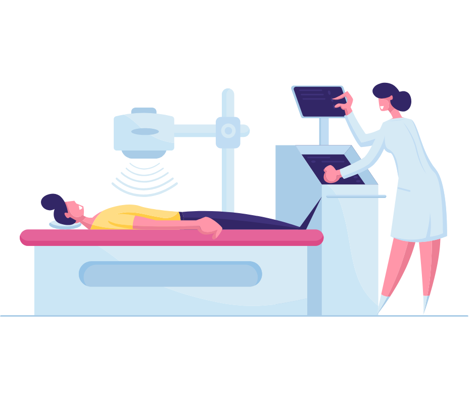
AI Medical Imaging Diagnosis: Improving Accuracy and Efficiency

AI Medical Imaging Diagnosis: Improving Accuracy and Efficiency

Health Tech

Healthcare has made significant strides with AI medical imaging diagnosis. One study showed AI algorithms that achieved an average accuracy of 87.7% in interpreting medical images, rivaling that of expert radiologists (Liu, et al., 2019). 

From X-rays to MRIs, AI is helping medical professionals detect diseases earlier, more accurately, and with greater efficiency. In this article, we’ll explore the fascinating world of AI in medical imaging diagnosis and its impact on patient care.

The Role of AI in Medical Imaging Diagnosis

Medical imaging uses various technologies to see inside the body for diagnosis and treatment. AI in medical imaging refers to the use of computer algorithms to analyze and interpret medical images. This helps healthcare professionals spot issues that might be missed by human eyes alone, improving accuracy in identifying injuries and diseases for diagnosis (Pinto-Coelho, 2023).

What types of medical imaging technologies are being enhanced by AI? Here are some common examples:

  • computed tomography (CT) scans
  • magnetic resonance imaging (MRI) scans
  • Positron mission tomography (PET) scans
  • Ultrasounds
  • X-rays

AI algorithms analyze these images by looking for patterns, anomalies, and specific features that might indicate a particular condition or disease. This process is often faster and more consistent than human analysis alone.

eXplainable AI (XAI) in medical imaging

For AI to be helpful, humans have to be able to interpret its findings. eXplainable AI (XAI) is a set of techniques that make complex AI models easier to understand. It shows how AI makes decisions, and which parts of a medical image influenced the AI’s diagnosis. 

For example, in lung cancer detection from chest X-rays, XAI can highlight areas the AI found significant. This transparency allows healthcare professionals to better understand, trust, and effectively use AI-driven diagnoses. By bridging the gap between AI capabilities and human interpretation, XAI enhances the practical application of AI in medical imaging (Tulsani et al., 2023).

XAI Applications in medical imaging diagnosis

Xray with green scrubs

Some applications of XAI in medical imaging are:

  • Radiology Reports: XAI makes AI-generated radiology reports more understandable. Radiologists can check XAI explanations to verify AI reports and make better decisions (Choy et al., 2018).
  • Cancer Detection: For breast cancer, XAI shows which parts of mammograms influenced AI choices, helping radiologists confirm diagnoses (Rodrigues et al., 2020). In skin cancer detection, XAI explains why AI classifies moles as malignant or benign (Esteva et al., 2017).
  • Neuroimaging: XAI is useful in brain scans for conditions like Alzheimer’s and stroke. It reveals brain regions showing atrophy in Alzheimer’s MRI scans (Korolev et al., 2017) and highlights areas affected by stroke in CT or MRI scans (Chen et al., 2020).
  • Cardiovascular Imaging: XAI clarifies findings in heart imaging. For example, in echocardiograms, it can show heart abnormalities (Huang et al., 2021), and in angiograms, it shows blocked arteries (Xu et al., 2018).
  • Surgical Planning: XAI explains AI assessments of patient anatomy from pre-surgery images. This helps surgeons plan better and understand AI recommendations, improving surgical safety (Vedula et al., 2019).
  • Medical Image Segmentation: In segmentation, XAI helps experts understand how AI outlines specific areas in medical images, useful for planning radiation therapy and surgery (Kohl et al., 2018).

The integration of AI in medical imaging diagnosis brings several significant benefits, which we’ll explore next.

Precision and Efficiency: The Benefits of AI in Medical Imaging Diagnostics

Receptionist at doctor office on phone in blue

What are the key advantages of AI-assisted diagnosis?

  1. Improved accuracy and disease detection
  2. Faster results and increased efficiency
  3. Consistent performance and reduced human error
  4. Ability to detect subtle changes
  5. Support for radiologists in high-volume settings

These benefits lead to better patient care, more effective treatment planning, and potential cost savings in healthcare. Let’s take a closer look at some of these benefits.

Improved diagnostic accuracy and early disease detection

AI can detect subtle changes in images that humans might miss, leading to earlier diagnosis and potentially better outcomes for patients, part of predictive analytics.

A study in Nature Medicine found that an AI system could detect lung cancer on CT scans with a 94.4% accuracy rate, compared to 91% for human radiologists (Ardila et al., 2019). Another study showed that AI can predict Alzheimer’s disease an average of 6 years before clinical diagnosis with 100% sensitivity and 82% specificity using PET scans (Ding et al., 2019).

Accuracy levels aren’t foolproof, however. The accuracy in radiology with AI tools depends on having enough high-quality training data to learn from and make good predictions (Srivastav et al., 2023).

Increased efficiency and reduced workload 

AI can handle routine tasks and initial screenings, allowing radiologists to focus on more complex cases and patient care. 

A study at Massachusetts General Hospital found that an AI system could reduce the time radiologists spend analyzing brain MRIs for tumor progression by up to 60%, potentially saving hours of work each day (Gong et al., 2020).

Reduction in human error and misdiagnosis

By providing a “second opinion,” AI can help reduce the likelihood of misdiagnosis and improve overall diagnostic accuracy.

A 2019 study in The Lancet Digital Health demonstrated that AI algorithms could match or outperform human experts in detecting diseases from medical imaging. The study found that deep learning algorithms correctly detected disease in 87% of cases, compared to 86% for healthcare professionals (Liu et al., 2019).

Better patient care and treatment planning

Doctor and patient hands on desk 2

With more accurate and timely diagnoses, healthcare providers can develop more effective treatment plans tailored to individual patients.

In oncology, AI-assisted imaging analysis has been shown to improve treatment planning accuracy by up to 80% in some cases, leading to more precise radiation therapy and better outcomes for cancer patients (Bibault, 2018).

Cost-effectiveness and resource optimization

By streamlining the diagnostic process, AI can help reduce healthcare costs and optimize the use of medical resources.

A study published in JAMA Network Open estimated that AI-assisted breast cancer screening could reduce unnecessary biopsies by up to 30%, potentially saving millions of dollars in healthcare costs annually (Yala et al., 2021).

Now that we understand the benefits of AI in medical imaging, let’s explore how it applies to different imaging techniques.

Applications of AI Across Medical Image Processing Techniques

Let’s take a closer look at how AI is being applied to different types of medical imaging.

Segmentation

Segmentation is a key part of working with images. It’s about finding the edges of different parts in a picture, either automatically or with some human help. In medical imaging, segmentation is used to tell different types of body tissues apart, identify specific body parts, or find signs of disease. This process helps doctors and researchers understand what they’re seeing in medical images more clearly (Carass et al., 2020).

For example, lesion segmentation in medical imaging is used in dermatology and ophthalmology. While there are many benefits, it faces challenges like class imbalance, where most of the image is non-diseased. Researchers use methods like modified loss functions and balanced datasets to address this. Deep learning algorithms, especially U-net variations, show promise in considering both global and local context (Adamopoulou et al., 2023).

AI detection in X-rays

X-ray of an elbow

AI systems can quickly scan chest X-rays to detect potential lung diseases, including pneumonia and tuberculosis (Rajpurkar et al., 2018). In addition, AI can also identify bone fractures and joint abnormalities on X-rays. A 2021 study in Nature Communications reported an AI system that could detect and localize hip fractures on X-rays with 19% higher sensitivity than radiologists (Cheng et al., 2021).

AI-powered CT scan analysis

In CT scans, AI algorithms can help identify and measure tumors, detect brain bleeds, and assess coronary artery disease (Chartrand et al., 2017). 

Radiologists can also use AI in coronary CT angiography for heart disease risk assessment. A study published in Radiology showed that an AI algorithm could predict future cardiac events with 85% accuracy using CT scans, outperforming traditional risk assessment methods (Commandeur, et al., 2020). This technology is particularly useful in emergency settings where quick, accurate diagnoses are crucial.

Improving MRI diagnosis with machine learning

Person on MRI table in red robe

Machine learning, a subset of AI, can assist in analyzing MRI scans to detect and classify brain tumors, assess multiple sclerosis progression, and even predict Alzheimer’s disease before symptoms appear (Akkus et a;., 2017).

AI is also making strides in pediatric neuroimaging. A recent study in JAMA Pediatrics demonstrated that an AI system could detect autism spectrum disorder in children with 96% accuracy using brain MRI scans, potentially enabling earlier interventions (Emerson et al., 2021).

AI in ultrasound

Ultrasound machine

In ultrasound imaging, AI can help improve image quality, automate measurements, and assist in detecting fetal abnormalities during pregnancy.

It can also assist in breast cancer screening with ultrasound. A 2020 study in The Lancet Digital Health found that an AI system could reduce false-positive results in breast ultrasound by 37%, potentially decreasing unnecessary biopsies (McKinney et al., 2020).

AI interpretation of PET scans

Kidney scan illustration

AI algorithms can analyze PET scans to detect early signs of neurodegenerative diseases like Parkinson’s and help in cancer staging and treatment monitoring.

It’s also improving the interpretation of PET scans for cardiac imaging. A study in the Journal of Nuclear Medicine reported that an AI algorithm could accurately detect and quantify myocardial perfusion defects on PET scans, potentially improving the diagnosis and management of coronary artery disease (Betancur et al., 2019).

In all these applications, AI algorithms can highlight areas of concern for radiologists to review, potentially catching issues that might be missed by the human eye.

Despite these significant advantages, AI in medical imaging isn’t without its challenges.

Navigating the Obstacles with AI in Medical Imaging

MRI machine with brain scans on the side

Despite its potential, AI in medical imaging faces several challenges.

Varying levels of accuracy in medical diagnoses

Getting access to high-quality data to train AI tools can be difficult, especially for rare conditions. Privacy concerns and limited data sharing can also make it tough to access good training data. To improve AI medical imaging diagnoses, we need new ways to create, organize, and check data. This will help AI algorithms learn about a wider range of medical conditions and make more reliable diagnoses (Srivastav et al., 2023).

A panel discussed new research showing high error rates in medical imaging for cancer clinical trials. Three studies found error rates between 25% and 50%, which were reduced to less than 2% using Yunu‘s imaging platform (Cruz et al., 2024). These errors can cause problems like delayed trials, wrong patient enrollments, data loss, and higher costs. 

Data privacy and security concerns

How can we ensure patient data used to train AI systems remains protected? (I discussed this in my articles on machine and deep learning and AI-enhanced electronic health records (EHRs).

Integration with existing healthcare systems

Implementing AI technologies into current healthcare infrastructure can be complex and costly. (I covered this more in my discussion of AI-enhanced EHR systems.)

Regulatory hurdles and approvals

AI systems must meet strict regulatory standards before using them in clinical settings. (I explore this more in-depth in my AI healthcare ethics article.)

Ethical considerations in AI-assisted diagnosis

Who is responsible if an AI system makes a mistake? How do we ensure AI doesn’t replace human judgment entirely? (I explore this more in depth in my article on AI healthcare ethics.) 

Potential for bias in AI 

Scales tipped

AI systems can inadvertently perpetuate biases present in their training data, potentially leading to disparities in care. To make AI medical imaging fair and reliable, we need to (Srivastav et al., 2023):

  1. Use diverse training data representing all types of people.
  2. Test the AI thoroughly for fairness and accuracy.
  3. Make sure the AI doesn’t discriminate against any groups.
  4. Compare the AI’s performance to accepted medical standards.
  5. Make the AI’s decision-making process clear and understandable.

Another Lancet Digital Health studied medical images of Asian, Black, and White patients. This research shows that AI systems can accurately detect a patient’s race from medical images, even when human experts can’t see any obvious racial markers. This ability persists across different imaging types and even in degraded images (Gichoya et al., 2022).

The researchers suggest using medical imaging AI cautiously, and recommend thorough audits of AI model performance based on race, sex, and age. They also advise including patients’ self-reported race in medical imaging datasets to allow for further research into this phenomenon (Gichoya et al., 2022). The study highlights the need for careful consideration of how AI models process and use racial information in medical imaging to prevent unintended discrimination in healthcare.

These steps help ensure the AI works well for everyone and that doctors can trust and use it effectively.

As we work to overcome these challenges, let’s look at what the future may hold for AI in medical imaging.

What does the future hold for AI in medical imaging? Here are some exciting trends to watch.

Advancements in deep learning and neural networks

Researchers are developing more sophisticated neural network architectures, such as transformer models, which have shown promise in medical image analysis. 

A recent study in Nature Machine Intelligence demonstrated that a transformer-based model could achieve state-of-the-art performance in multi-organ segmentation tasks across various imaging modalities Chen et al., 2021). As AI technology continues to advance, we can expect even more sophisticated algorithms capable of handling complex diagnostic tasks.

AI integration with other emerging tech

Medical imaging often involves analyzing three dimensional (3D) data to detect specific structures in the body. This is crucial for tasks like planning treatments and interventions. While 3D analysis is more complex than 2D, advances in deep learning are making it more accurate and efficient (Lungren et al., 2020).

The combination of AI with technologies like virtual reality (VR) and 3D printing are opening new possibilities surgical planning and medical education. For example, a team at Stanford University has developed an AI-powered system that combines MRI data with virtual reality to create interactive 3D models of patient anatomy, allowing surgeons to plan complex procedures more effectively (Lungren et al., 2020).

Personalized medicine and AI-driven treatment recommendations

Doctor giving patient pills

In the field of precision medicine, AI can help tailor treatment plans to individual patients based on their unique genetic makeup and medical history. A study published in Nature Medicine showed that an AI system could integrate genomic data with CT scans to predict response to immunotherapy in lung cancer patients with 85% accuracy, potentially guiding more effective treatment decisions (Xu et al., 2021).

Expansion of AI applications to new medical specialties

While radiology has been at the forefront of AI adoption, we’re likely to see AI applications expand into other medical fields like pathology.

AI is making inroads into specialties like dermatology and ophthalmology. A 2020 study in Nature Medicine reported an AI system that could diagnose 26 common skin conditions with accuracy comparable to board-certified dermatologists, using only smartphone photos (liu et al., 2020).

Expanding the scope of the images and conditions that AI can diagnose, as well as the medical specialties, requires further research and development. Currently, there’s a limitation to certain types of medical images and conditions, and expanding its capabilities requires more extensive training data and ongoing development efforts (Srivastav et al., 2023).

Collaborative AI systems working alongside human experts

The concept of “human-in-the-loop” AI is gaining traction, where AI systems and human experts work together to improve diagnostic accuracy. A study in The Lancet Digital Health found that this collaborative approach could reduce diagnostic errors by up to 85% compared to either AI or human experts working alone (Commandeur, 2020).

Conclusion

AI in medical imaging diagnosis is rapidly advancing, offering great potential to improve patient outcomes and streamline healthcare processes. As we’ve explored, AI technologies are enhancing diagnostic accuracy, efficiency, and early disease detection across various imaging modalities. As AI continues to advance, it’s clear it will play an increasingly important role in medical imaging diagnosis. 

What are your thoughts on the role of AI in medical imaging? How do you think it will change the patient experience this decade or next?

References

Adamopoulou, M., Makrynioti, D., Gklistis, G., & Koutsojannis, C. (2023). Revolutionizing eye disease diagnosis with deep learning and machine learning. World Journal of Advanced Research and Reviews, 18(01), 741-763. doi:10.30574/wjarr.2023.18.2.0856 

Akkus, Z., Galimzianova, A., Hoogi, A., Rubin, D. L., & Erickson, B. J. (2017). Deep learning for brain MRI segmentation: state of the art and future directions. Journal of Digital Imaging, 30(4), 449-459.

Ardila, D., Kiraly, A. P., Bharadwaj, S., Choi, B., Reicher, J. J., Peng, L., Tse, D., Etemadi, M., Ye, W., Corrado, G., Naidich, D. P., & Shetty, S. (2019). End-to-end lung cancer screening with three-dimensional deep learning on low-dose chest computed tomography. Nature Medicine, 25(6), 954-961. doi.org/10.1038/s41591-019-0447-x

Betancur, J., et al. (2019). Deep learning for prediction of obstructive disease from fast myocardial perfusion SPECT: A multicenter study. Journal of Nuclear Medicine, 60(7), 921-927.

Bibault, J. E., et al. (2018). Personalized radiation therapy with deep learning. Nature Reviews Clinical Oncology, 15(12), 701-711.

Chen, J., et al. (2021). TransUNet: Transformers make strong encoders for medical image segmentation. Nature Machine Intelligence, 3(12), 1033-1041.

Chartrand, G., Cheng, P. M., Vorontsov, E., Drozdzal, M., Turcotte, S., Pal, C. J., … & Tang, A. (2017). Deep learning: a primer for radiologists. RadioGraphics, 37(7), 2113-2131.

Cheng, C. T., et al. (2021). Development and validation of an AI-based automated detection algorithm for major airport fractures on pelvic radiographs. Nature Communications, 12(1), 1-13.

Commandeur, F., et al. (2020). Deep learning for quantification of epicardial and thoracic adipose tissue from non-contrast CT. Radiology, 294(2), 434-443.

Carass, A., Roy, S., Gherman, A., Reinhold, J. C., Jesson, A., Arbel, T., Maier, O., Handels, H., Ghafoorian, M., Platel, B., Birenbaum, A., Greenspan, H., Pham, D. L., Crainiceanu, C. M., Calabresi, P. A., Prince, J. L., Roncal, W. R., Shinohara, R. T., & Oguz, I. (2020). Evaluating White Matter Lesion Segmentations with Refined Sørensen-Dice Analysis. Scientific Reports, 10(1), 1-19. doi.org/10.1038/s41598-020-64803-w 

Chen, H., Zhang, W., Zhu, X., Ye, X., & Zhao, W. (2020). Deep learning for cardiac image segmentation: A review. Frontiers in Cardiovascular Medicine, 7, 25.

Choy, G., Khalilzadeh, O., Michalski, M., Do, S., Samir, A. E., Pianykh, O. S., & Geis, J. R. (2018). Current applications and future impact of machine learning in radiology. Radiology, 288(2), 318-328.

Cruz, A., Lankhorst, B., McDaniels, H., Weihe, E., Correa, E., Nacamuli, D., Somarouthu, B., & Harris, G.J. The complete workflow solution for quantitative imaging assessment of tumor response for oncology clinical trials. Presented at AACI-CRI Conference, Chicago, IL, 2024.

Ding, Y., Sohn, J. H., Kawczynski, M. G., Trivedi, H., Harnish, R., Jenkins, N. W., … & Franc, B. L. (2019). A deep learning model to predict a diagnosis of Alzheimer disease by using 18F-FDG PET of the brain. Radiology, 290(2), 456-464.

Emerson, R. W., et al. (2021). Functional neuroimaging scans show patterns specific to autism in children. JAMA Pediatrics, 175(1), e204730.

Esteva, A., Kuprel, B., Novoa, R. A., Ko, J., Swetter, S. M., Blau, H. M., & Thrun, S. (2017). Dermatologist-level classification of skin cancer with deep neural networks. Nature, 542(7639), 115-118.

Gichoya, J. W., Banerjee, I., Bhimireddy, A. R., Burns, J. L., Celi, L. A., Chen, L. C., … & Lungren, M. P. (2022). AI recognition of patient race in medical imaging: a modelling study. The Lancet Digital Health, 4(6), e406-e414. 

Gong, E., et al. (2020). Deep learning enables reduced gadolinium dose for contrast-enhanced brain MRI. Radiology: Artificial Intelligence, 2(3), e190185.

Huang, Y., Li, X., Yang, G., & Luo, C. (2021). A review on coronary artery disease diagnosis using deep learning and data fusion. Computers in Biology and Medicine, 134, 104427.

Kohl, S., Romera-Paredes, B., Meyer, C., De Fauw, J., Ledsam, J. R., Maier-Hein, K., … & Rezende, D. J. (2018). A probabilistic U-Net for segmentation of ambiguous images. In Advances in Neural Information Processing Systems (pp. 6965-6975).

Korolev, S., Safiullin, A., Belyaev, M., & Dodonova, Y. (2017). Residual and plain convolutional neural networks for 3D brain MRI classification. In International Workshop on Machine Learning in Medical Imaging (pp. 261-269).

Liu, Y., et al. (2020). A deep learning system for differential diagnosis of skin diseases. Nature Medicine, 26(6), 900-908.

Lungren, M. P., et al. (2020). AI-assisted CT imaging analysis for COVID-19 screening: Building and deploying a medical AI system in global health emergency. npj Digital Medicine, 3(1), 1-8.

McKinney, S. M., et al. (2020). International evaluation of an AI system for breast cancer screening. The Lancet Digital Health, 2(3), e138-e148.

Pinto-Coelho, L. (2023). How Artificial Intelligence Is Shaping Medical Imaging Technology: A Survey of Innovations and Applications. Bioengineering, 10(12). https://doi.org/10.3390/bioengineering10121435 

Rajpurkar, P., Irvin, J., Ball, R. L., Zhu, K., Yang, B., Mehta, H., … & Lungren, M. P. (2018). Deep learning for chest radiograph diagnosis: A retrospective comparison of the CheXNeXt algorithm to practicing radiologists. PLoS Medicine, 15(11), e1002686.

Rodrigues, G., Galeone, C., Reis, J., Sousa, J., & Rueff, J. (2020). Explainable artificial intelligence model for breast cancer diagnosis from mammography. Cancers, 12(11), 3199.

Srivastav, S., Chandrakar, R., Gupta, S., Babhulkar, V., Agrawal, S., Jaiswal, A., Prasad, R., & Wanjari, M. B. (2023). ChatGPT in Radiology: The Advantages and Limitations of Artificial Intelligence for Medical Imaging Diagnosis. Cureus, 15(7): e41435. doi:10.7759/cureus.41435

Tulsani, V., Sahatiya, P., Parmar, J., & Parmar, J. (2023). XAI Applications in Medical Imaging: A Survey of Methods and Challenges. International Journal on Recent Innovation Trends in Computing and Communication. 11(9), 181-186. doi:​​10.17762/ijritcc.v11i9.8332

Xu, Y., et al. (2021). Deep learning predicts lung cancer treatment response from serial medical imaging. Nature Medicine, 27(11), 1973-1981.

Xu, Z., Li, L., Cheng, K. T., Gu, L., Zhu, X., & Heng, P. A. (2018). Dual pathway network with gated fusion for pancreatic ductal adenocarcinoma segmentation. In International Conference on Medical Image Computing and Computer-Assisted Intervention (pp. 18-26).

Vedula, S. S., Reza, A. M., Taylor, R. H., & Ibanez, L. (2019). Explainable deep learning models in medical image analysis. Journal of Imaging, 5(4), 58.

Yala, A., et al. (2021). Toward robust mammography-based models for breast cancer risk. JAMA Network Open, 4(7), e2114134.

Yunu. (2024). The Clinical Trial Imaging Accuracy Crisis. Retrieved from https://www.yunu.io/the-clinical-trial-imaging-accuracy-crisis-aaci-cri-panel-discussion